| Home | Sources Directory | News Releases | Calendar | Articles | | Contact | |
Lysosome


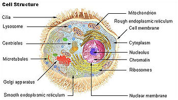
(1) nucleolus
(2) nucleus
(3) ribosomes (little dots)
(4) vesicle
(5) rough endoplasmic reticulum (ER)
(6) Golgi apparatus
(7) Cytoskeleton
(8) smooth endoplasmic reticulum
(9) mitochondria
(10) vacuole
(11) cytosol
(12) lysosome
(13) centrioles within centrosome
Lysosomes are spherical organelles that contain enzymes (acid hydrolases) that break up endocytized materials and cellular debris. They are found in animal cells, while in yeast and plants the same roles are performed by lytic vacuoles.[1] Lysosomes digest excess or worn-out organelles, food particles, and engulfed viruses or bacteria. The membrane around a lysosome allows the digestive enzymes to work at the 4.5 pH they require. Lysosomes fuse with vacuoles and dispense their enzymes into the vacuoles, digesting their contents. They are created by the addition of hydrolytic enzymes to early endosomes from the Golgi apparatus. The name lysosome derives from the Greek words lysis, which means to separate; and soma, which means body. They are frequently nicknamed "suicide-bags" or "suicide-sacs" by cell biologists due to their role in autolysis. Lysosomes were discovered by the Belgian cytologist Christian de Duve in 1949.
The size of lysosomes varies from 0.1'1.2 î�m.[2] At pH 4.8, the interior of the lysosomes is acidic compared to the slightly alkaline cytosol (pH 7.2). The lysosome maintains this pH differential by pumping protons (H+ ions) from the cytosol across the membrane via proton pumps and chloride ion channels. The lysosomal membrane protects the cytosol, and therefore the rest of the cell, from the degradative enzymes within the lysosome. The cell is additionally protected from any lysosomal acid hydrolases that leak into the cytosol as these enzymes are pH-sensitive and function less well in the alkaline environment of the cytosol.
Contents |
[edit] Enzymes
Some important enzymes found within lysosomes include:
- Lipase, which digests lipids
- Amylase, which digest carbohydrates (e.g., sugars)
- Proteases, which digest proteins
- Nucleases, which digest nucleic acids
- phosphoric acid monoesters.
Lysosomal enzymes are synthesized in the cytosol and the endoplasmic reticulum, where they receive a mannose-6-phosphate tag that targets them for the lysosome[citation needed] . Aberrant lysosomal targeting causes inclusion-cell disease, whereby enzymes do not properly reach the lysosome, resulting in accumulation of waste within these organelles.[citation needed]
[edit] Functions
Lysosomes are the cell's garbage disposal system. They are used for the digestion of macromolecules from phagocytosis (ingestion of other dying cells or larger extracellular material, like foreign invading microbes), endocytosis (where receptor proteins are recycled from the cell surface), and autophagy (where in old or unneeded organelles or proteins, or microbes that have invaded the cytoplasm are delivered to the lysosome). Autophagy may also lead to autophagic cell death, a form of programmed self-destruction, or autolysis, of the cell, which means that the cell is digesting itself.
Other functions include digesting foreign bacteria (or other forms of waste) that invade a cell and helping repair damage to the plasma membrane by serving as a membrane patch, sealing the wound. In the past, lysosomes were thought to kill cells that were no longer wanted, such as those in the tails of tadpoles or in the web from the fingers of a 3- to 6-month-old fetus. While lysosomes digest some materials in this process, it is actually accomplished through programmed cell death, called apoptosis.[3][4]
[edit] Clinical relevance
There are a number of lysosomal storage diseases that are caused by the malfunction of the lysosomes or one of their digestive proteins; examples include Tay-Sachs disease and Pompe's disease. These diseases are caused by a defective or missing digestive protein, which leads to the accumulation of substrates within the cell, impairing metabolism.
In the broad sense, these can be classified as mucopolysaccharidoses, GM2 gangliosidoses, lipid storage disorders, glycoproteinoses, mucolipidoses, or leukodystrophies.
|
Proteins in different cellular compartments and structures tagged with green fluorescent protein. |
[edit] External links
- 3D structures of proteins associated with lysosome membrane
- Hide and Seek Foundation For Lysosomal Research Team
- Self-Destructive Behavior in Cells May Hold Key to a Longer Life
- Mutations in the Lysosomal Enzyme'Targeting Pathway and Persistent Stuttering
[edit] References
- ^ Samaj J, Read ND, Volkmann D, Menzel D, Baluska F (August 2005). "The endocytic network in plants". Trends Cell Biol. 15 (8): 425'33. doi:10.1016/j.tcb.2005.06.006. PMID 16006126.
- ^ Kuehnel, W (2003). Color Atlas of Cytology, Histology, & Microscopic Anatomy (4th ed.). Thieme. pp. 34. ISBN 1-58890-175-0.
- ^ Lysosomes and Peroxisomes
- ^ Mader, Sylvia. (2007). Biology 9th ed. McGraw Hill. New York. ISBN 978-0072464634
 This article incorporates public domain material from the NCBI document "Science Primer".
|
|||||||||||||||||
|
SOURCES.COM is an online portal and directory for journalists, news media, researchers and anyone seeking experts, spokespersons, and reliable information resources. Use SOURCES.COM to find experts, media contacts, news releases, background information, scientists, officials, speakers, newsmakers, spokespeople, talk show guests, story ideas, research studies, databases, universities, associations and NGOs, businesses, government spokespeople. Indexing and search applications by Ulli Diemer and Chris DeFreitas.
For information about being included in SOURCES as a expert or spokesperson see the FAQ . For partnerships, content and applications, and domain name opportunities contact us.
